Monthly Archives: October 2017

Spinal Cord Injury May Increase the Risk of Alzheimer’s Disease

It may be surprising to some people to learn that physical injuries can contribute to neurodegenerative diseases. In mice, traumatic brain injuries have been previously shown to induce an Alzheimer’s-like condition, complete with amyloid plaques and neuroinflammation. In a study published this week in the journal Spinal Cord, researchers from the National Taiwan University investigated whether a similar connection exists between spinal cord injuries (SCI) and Alzheimer’s disease.

The researchers utilized medical records from Taiwan’s National Health Insurance Research Database. Their analyses included 9,257 individuals with an SCI and 37,028 non-SCI individuals, with an average age of approximately 63 years. When selecting the subjects for the study, the researchers applied an algorithm that would correct for the effects of other Alzheimer’s disease risk factors, such as age, sex, and cardiovascular health.

Over the course of a three-year period, a total of 25 SCI inviduals and 57 non-SCI individuals were diagnosed with Alzheimer’s. These numbers are quite low, possible due to the difficulty in diagnosing Alzheimer’s with high certainty, since its symptoms are similar to other forms of dementia. Cumulatively, the incidence of an Alzheimer’s diagnosis during the three-year study period was 71% higher in people with a SCI compared to people without a SCI. (I want to emphasize that this does not mean SCI patients are 71% more likely to get Alzheimer’s over their entire lifetime; this number only applies to the three-year period examined in this study.)

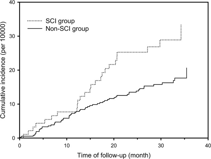
This figure from the paper shows the incidence of Alzheimer’s disease over a three-year period. The SCI individuals had an increased risk of Alzheimer’s than the non-SCI individuals.

This study is the first large-scale, longitudinal analysis to demonstrate a correlation between SCI and Alzeimer’s disease. Future research is warranted to determine what might be causing this connection. The authors suggested several possible explanations. It has been previously shown that tau and the amyloid precursor protein are deposited throughout the spinal cord following a SCI. These proteins are both closely linked to the pathogenesis of Alzheimer’s disease, so this could be a possible disease mechanism. Another possibility is that the widespread inflammation triggered by a SCI could perturb the delicate chemistry of molecules within the brain.

There are several important caveats to note with this study. For one thing, while the researchers accounted for health-related Alzheimer’s risk factors, including various medical conditions like diabetes and stroke, their records did not include information about lifestyle-related risk factors, such as smoking or exercise. These factors could have potentially skewed the analysis. Also, since the follow-up period was only three years, the data does not give us information about any longer-term effects of SCI. Finally, while the total sample size was large, only a small subset of subjects were diagnosed with Alzheimer’s, reducing the statistical power of the analysis. Future studies will need to address these problems in order to provide further insight into the emerging connection between SCI and Alzheimer’s disease.


Enjoy this post? Help it to grow by sharing on social media!
Want more? Follow AlzScience via email, Facebook, or Twitter!

Study Shows How Alzheimer’s Affects Men’s and Women’s Brains Differently

Women are around twice as likely to develop Alzheimer’s disease during their lifetimes compared to men. This effect is also seen in mouse models of the disease. When mice are genetically engineered to developed Alzheimer’s, the female mice tend to have an earlier diseae onset and more severe pathology compared to hte male mice. The reasons for this discrepancy are unknown. However, a recent study published in Neurobiology of Aging attempted to shed some light on the mystery.

The researchers were interested in studying adult neurogenesis, which is our brains’ ability to create new neurons throughout our lives. Neurogenesis primarily occurs in two areas of the brain: the olfactory bulb, which is involved with the sense of smell, and the hippocampus, which is important for memory. In this study, the scientists wanted to figure out whether neurogenesis in the hippocampus was different for male and female mice with Alzheimer’s disease.

First, they subjected the mice to a test designed to test spatial memory, which is particularly important for the hippocampus. They placed the mice in a container with two objects and allowed them to explore for a few minutes. The next day, they placed the mice back in the container, but now one of the objects had been moved to a new location. We would expect the mice to spend more time sniffing and investigating the object that had been moved, and less time sniffing the object that was in the same place as before.

Screenshot 2017-10-13 12.44.30.png

When one of the cups is moved to a new location, the mouse should spend more time sniffing it compared to the cup that wasn’t moved. This test is used to analyze the mouse’s spatial memory. Image Source

The result was interesting. While the male mice had no trouble rembering which cup had been moved, the female mice did significantly worse, spending the same amount of time sniffing both of the cups. This suggested that the female mice might have some impairment in their spatial memory.

Next, the researches looked at the mice’s brains. They used a technique that caused all newly-born neurons to be labeled bright green, and counted how many neurons were born in the hippocampus during a two-week period. The male mice produced more than four times as many neurons as the female mice. Conversely, the female mice had nearly twice as many astrocytes in their hippocampi compared to the males. Astrocytes are another type of brain cell that is often associated with inflammation. These results suggested that the female mice’s brains were producing many astrocytes but few neurons, perhaps contributing to their impaired spatial memory.

Screenshot 2017-10-13 12.54.35.png

This image from the paper shows astrocytes labeled in red. In the top right panel, you can see that the female Alzheimer’s (APP/PS1) mice have far more red area than the males, indicating a greater number of astrocytes.

The results of this study suggest that the brains of female mice with Alzheimer’s may be devoting so many resources to creating new astrocytes that there’s not enough left to create neurons. However, it opens up many new questions. What is causing this overproliferation of astrocytes in the female mice? The authors of the paper suggest estrogen as a possible cause, since this hormone has been shown to influence memory. Additional studies are needed to determine the true cause.


Enjoy this post? Help it to grow by sharing on social media!
Want more? Follow AlzScience via email, Facebook, or Twitter!